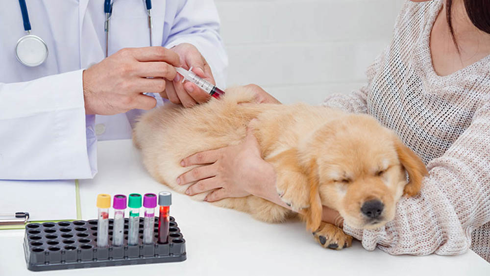
How Do Dogs Get X-Rays?
X-rays are one of the best ways for your vet to see what’s going on inside your dog’s body. And there are plenty of conditions the place an X-ray is usually a highly effective diagnostic device. Here are a number of the commonest causes your dog might have an X-ray. If your vet has beneficial an X-ray for your dog, you’re likely wondering how much it'll value and what to expect with the process.
Reimbursement for Vet Bill After Payment
X-rays are best used to evaluate bones and joints, as properly as the chest and stomach cavities. They are significantly useful for detecting fractures, bone abnormalities, and adjustments within the dimension and form of organs. The resulting images will present the form and structure of the bones within the tail, in addition to any abnormalities, such as fractures or tumors. Hip dysplasia is a typical situation in canine, significantly in large breeds, the place the hip joint doesn’t form correctly. It may cause pain, lameness, and arthritis, and can be diagnosed with an x-ray. The canine might want to lie still during the process, and the vet will take a number of pictures from totally different angles to get a comprehensive view of the organs.
X-Rays For Pregnant Dogs
You’ll have peace of mind understanding you'll have the ability to manage sudden vet payments with out breaking the financial institution. There are all kinds of distinct aspects that might affect the fee. Be prepared to pay an additional price, for example, in case your canine companion needs to be anesthetized for the x-ray. The worth may also differ relying on the location of the x-ray machine or on the breed of the dog. The cost of X-rays for canine varies, but pet dad and mom can anticipate to pay anyplace from $200 to $500 or more, notably if sedation, common anesthesia, or additional photographs are needed.
How Much Do Dog X-Rays Cost?
With protection, you’ll usually pay less out-of-pocket for these important diagnostic exams, easing financial strain throughout emergencies. On the downside, publicity to X-rays poses potential hazards similar to the danger of tissue harm and the development of radiation-induced health issues over time. While not all canine require sedation for an X-ray, sedation may help shorten the exposure time by reducing the dog’s motion throughout imaging. This helps forestall blurry or distorted pictures, which could otherwise necessitate additional X-rays and radiation publicity.
 Si observas alguno de estos síntomas en tu perro, es esencial buscar atención veterinaria inmediatamente, puesto que el edema pulmonar puede ser una urgencia médica que requiere tratamiento urgente. Las infecciones pulmonares, como la neumonía, pueden causar inflamación en los pulmones y la acumulación de líquido. Las alergias asimismo tienen la posibilidad de desencadenar una respuesta inflamatoria en los pulmones, lo que transporta a la capacitación de edema. Además, ciertos medicamentos tienen la posibilidad de causar una reacción adversa en el sistema respiratorio del perro, lo que resulta en edema pulmonar. En el presente artículo, exploraremos en detalle qué es el edema pulmonar, los signos y síntomas que tienen la posibilidad de señalar su presencia, Https://Lomofy.Com.Ng y las medidas de tratamiento disponibles. Entender esta afección y de qué manera accionar en caso de urgencia es fundamental para garantizar la salud y bienestar de tu perro.
¿Puede el edema pulmonar provocar la muerte del animal?
Los análisis de sangre tienen la posibilidad de proveer información sobre la función renal, los escenarios de electrolitos y otros parámetros que pueden estar relacionados con la causa subyacente del edema pulmonar en el perro. Para la evaluación y diagnóstico del edema pulmonar en perros se efectuará una analítica sanguínea y una radiografía de tórax. Asimismo se puede monitorizar al perro (vigilar cambios en el segmento S-T que indican la oxigenación miocárdica).Otros factores a controlar son el PCV, la orina producida, la presión venosa central y el peso del animal. Si tu perro tiene una enfermedad cardiaca o renal, es esencial seguir las recomendaciones del veterinario para controlar la condición y achicar el peligro de edema pulmonar. En función de la cantidad de líquido y del resto del cuadro clínico, su gravedad va a ser mayor o menor y de esto va a depender la dificultad respiratoria que genere. El edema pulmonar en perros no se puede prevenir, pero se puede advertir a tiempo y tratar sin dificultades si observas el estado de tu mascota y acudes al veterinario lo antes posible.
Each sonographer develops his or her own system of completely evaluating the abdomen. Systematic analysis ensures that all buildings are scanned. The probe can be affixed to the animal's paw or tail during an anesthesia or during intensive care. The veterinarian will hear a sound signal reflecting coronary heart price and pulse. Doppler strategies use a 10-MHz ultrasound probe to detect blood move in an artery. Doppler sounds become audible when stress within the cuff is less than that within the artery. All noninvasive blood pressure monitors are technically easy to make use of; the equipment isn't cost-prohibitive and is available.
Veterinary doppler
A narrow beam of sound is projected into the guts, and the echo pattern and energy are displayed onto a persistence display, with the x-axis of the display representing time (y-axis is depth), similar to the familiar format of an ECG. The sample and amplitude of movement of the partitions of the chambers of the guts and valves may be evaluated, in addition to the scale of the respective buildings alongside the path of the sound beam. The M-mode format has very excessive temporal decision and thus is particularly suited to analysis of rapidly shifting constructions such as coronary heart valve leaflets. Considerable expertise is required to obtain and interpret diagnostic research. The M-mode examination has been coupled with real-time B-mode studies to improve the accuracy of beam placement and add additional information, similar to shape of the chamber. When used with a sphygmomanometer and an animal blood strain cuff of the proper dimension, systolicblood strain may be decided.
Covid-19 Products
Ultrasonography can additionally be used to direct biopsy devices to acquire tissue for a selected pathologic diagnosis, and is much safer and diagnostic than blind biopsy. This obviates the need for an open surgical exploration in plenty of circumstances. Lesions buried inside large organs such because the liver and kidneys which may not be detectable at surgical procedure could also be detected and biopsied with ultrasonographic steering. Presurgical analysis permits extra thorough and specific planning of surgical procedures and presurgical therapy of lesions. These procedures can frequently be safely performed beneath heavy sedation and analgesia. Ultrasound-guided biopsy and aspiration of lesions can be carried out in large animals with out the necessity for common anesthesia.
Vet-Dop 2 Doppler Blood Pressure System
In small animals, soft-tissue lesions of the ligaments, tendons, joint capsule, and articular cartilage of the shoulder and stifle joints are readily detectable by an skilled examiner. Most joints and muscle tissue may be evaluated by ultrasonography if the operator is acquainted with the normal anatomy and the style during which pathology of these structures is manifest on the image. The Dinamap is technically easier to use than the Doppler, for the explanation that precise location of the artery doesn't have to be found. It could be set to repeatedly measure the blood strain at an outlined interval (for instance, every 30 minutes) and will retain several readings till the machine is turned off.
The quantity by which the frequency is shifted is proportional to the velocity of the RBCs; whether or not it is a optimistic or unfavorable frequency shift is used to determine blood flow path. This is used to establish valvular regurgitation (insufficiency), elevated flow velocity (as in stenosis), or abnormal motion of the blood in the coronary heart or vessels elsewhere in the body. The most familiar one (and análise Laboratório veterinário the one that creates the precise picture of anatomy) is B-mode grayscale scanning. The sound beam is produced by a transducer positioned in touch with and acoustically coupled via a transmission gel to the animal.


